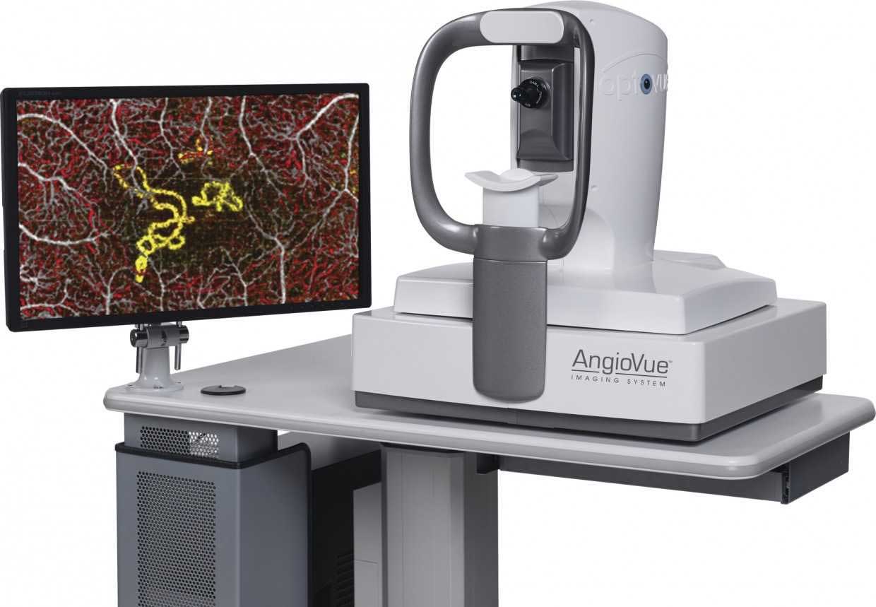
OPTICAL COHERENCE TOMOGRAPHY
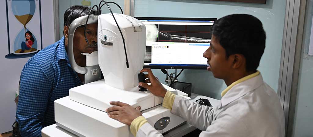
Optical coherence tomography (OCT) is a non-invasive imaging test that uses light waves to capture cross-section pictures of the retina and measure its layers for diagnosis and treatment guidance.
Optical coherence tomography (OCT) is a non-invasive imaging test that uses light waves to take cross-section pictures of the retina, the light-sensitive tissue lining the back of the eye.
With OCT, each of the retina's distinctive layers can be seen, allowing their thickness to be mapped and measured. These measurements help with diagnosis and provide treatment guidance for glaucoma and retinal diseases, such as age-related macular degeneration and diabetic eye disease.
To prepare a patient for an optical coherence tomography (OCT) exam, we may put dilating eye drops in the patient's eyes to widen the pupil and make it easier to examine the retina.
The patient will be seated in front of the OCT machine and will rest his or her head on a support to keep it motionless. The equipment will then scan the eye without touching it.
Scanning takes about 5 to 10 minutes. If the eyes were dilated, the patient may be sensitive to light for several hours after the exam.
OCT helps assess conditions such as: