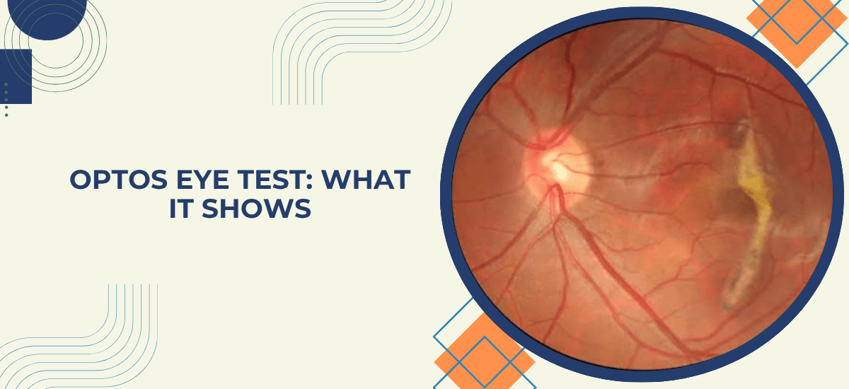
An Optos eye test is a retinal scan that captures up to roughly 200 degrees of the retina in a single image, where a standard retinal camera covers about 30 to 50 degrees. It uses a low-power scanning laser, involves no contact with the eye, and in many cases needs no dilating drops. Doctors rely on it to check for diabetic retinopathy, retinal tears and detachment, macular degeneration and other conditions that begin in the far edge of the retina, a region that routine examination can struggle to show in full.
Most people meet the Optos scan as a quick extra step in an eye check, and for many the image simply goes on file as a baseline. It matters more when the picture shows something at the outer edge, such as a thin patch or a small tear, because that area is hard to inspect any other way and problems there can develop without symptoms. Questions about what an image means are best answered by a retina specialist at an Eye Hospital in Mumbai.
“The retina does not hurt when something goes wrong at its edge. Tears and thin areas in the periphery can sit there for years without a single symptom, and that is exactly where a wide image helps. I would rather find a small problem on a scan than meet it later as a detachment.” — Dr. Vaishal Kenia, Chairman and Medical Director, Kenia Eye Hospital
What Is an Optos Eye Test?
The Optos test, also called an Optos scan, is a type of ultra-widefield retinal imaging. The device scans the retina with a low-power laser and builds a digital picture, so the result is one wide image rather than a set of small overlapping photographs. A single scan can show:
- the macula, which handles sharp central vision
- the optic nerve and the main blood vessels
- a large share of the peripheral retina, where tears and thin areas tend to form
Because the image is stored digitally, it can be compared year after year, which makes small changes easier to spot.
Wondering whether a retinal scan is right for you? A specialist can advise after a short consultation. Book an Appointment
What Happens During an Optos Eye Test?
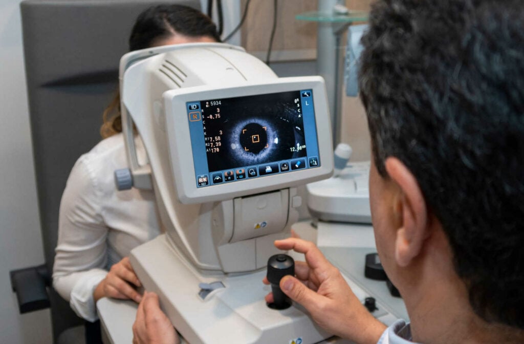
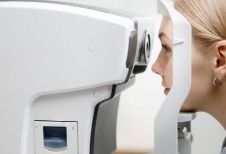
The test is quick and needs no preparation. You sit at the device, rest your chin and forehead on the support, and look at a light inside the machine. A flash follows, the image is captured in a fraction of a second, and the same is repeated for the other eye.
Nothing touches the eye, the scan uses no X-rays, and the whole test takes a few minutes. Some patients are still dilated afterwards if the specialist wants a closer look at a particular spot, but the scan itself does not depend on it.
What Does an Optos Scan Show?
The wide view helps most with conditions that start in the periphery or spread across the retina.
- Diabetic retinopathy. Leaking vessels and small bleeds in the outer retina can be missed on a smaller image. Our guide on diabetic retinopathy stages and treatment explains why regular screening matters.
- Retinal tears, holes and lattice degeneration, the weak spots that can lead to detachment, are often visible before they cause any symptoms.
- Retinal detachment, where the scan helps map how far it extends. See our guide on retinal detachment warning signs.
- Macular degeneration and retinal vein blockage, both of which leave visible changes across the retina.
- Changes at the optic nerve, along with moles, inflammation or growths in the retina.
When a weak spot is found early, it can often be sealed with laser, as described in our guide on barrage laser surgery.
Optos Scan vs Dilated Eye Exam: What Is the Difference?
Both look at the retina, but they work differently, and they often complement each other.
| Feature | Optos Scan | Dilated Eye Exam |
|---|---|---|
| Retinal area captured | Up to about 200 degrees in one image | Smaller view, built up by moving around the retina |
| Dilating drops | Often not needed | Needed |
| Time and after-effects | A few minutes, no lasting blur | Longer, with blurred vision for some hours |
| Permanent image record | Yes, easy to compare over time | Only if photographs are taken separately |
| Depth of the retina | Not shown, which needs an OCT scan | Specialist can judge depth through the lens |
An Optos scan does not replace a dilated exam in every case. The specialist decides which is needed, and many patients have both.
Who Should Get an Optos Eye Test?
A wide retinal image is most useful for people with a higher risk of retinal problems.
- People with diabetes, as part of yearly retina screening
- High myopia, since a stretched eye makes the retina thinner and more prone to tears
- Anyone with new floaters or flashes of light, to check for a tear
- A family history of retinal detachment or retinal disease
- Children, or others who find a standard exam difficult to sit through
- Anyone whose specialist wants a record to track changes over time
Kenia's retina clinic in Mumbai uses retinal imaging alongside OCT and fluorescein angiography, so the findings are read together rather than from one picture.
Why Choose Kenia Eye Hospital for an Optos Eye Test?
A wide image is only as useful as the person reading it. Kenia Eye Hospital has more than 26 years of experience in retinal care, and an Optos scan is read by the same specialists who treat the conditions it detects, so a finding on the image leads straight to a plan, whether that is monitoring, laser or injections. That joined-up approach is part of why Kenia is counted among the best eye hospital in Mumbai choices for retinal screening.
Due for a retina check, or advised to get a retinal scan? Book it with a specialist. Book an Appointment or call +91 75064 99962
