Understanding the DSAEK Cornea
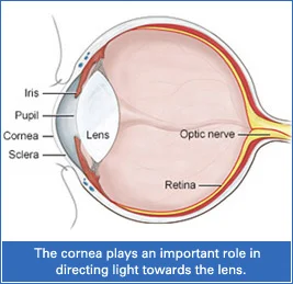
The cornea is the clear, front part of the eye that works like a window, allowing light to enter. It is responsible for about two-thirds of the eye’s focusing power, more than the eye’s natural lens.
A healthy cornea is transparent and gets its nutrients from the tear film. It should be free from blood vessels or cloudiness. However, the cornea can become damaged due to injury or disease, leading to scarring or clouding that affects your vision.
When the cornea becomes too cloudy or irregular, a corneal transplant may be needed. One advanced and less invasive option is called DSAEK (Descemet’s Stripping Automated Endothelial Keratoplasty), where only the damaged inner layer of the cornea is replaced with healthy donor tissue.
DSAEK offers a faster recovery and fewer complications compared to full-thickness cornea transplants. It also helps preserve more of your natural eye structure.
If you're considering treatment, Kenia Eye Hospital offers cornea transplant surgery in Mumbai using the latest techniques like DSAEK to restore clear vision.
For those dealing with corneal issues, it’s important to consult a skilled cornea specialist in Mumbai. Our team at Kenia Eye Hospital is equipped to guide you with expert care and advanced procedures.
WHO IS A CANDIDATE FOR DSAEK SURGERY?
DSAEK is a modern alternative to full cornea transplant surgery, but it’s not suitable for everyone. It works best for people who have damage only in the inner layer of the cornea, called the endothelium.
If the outer layers of the cornea are healthy but the inner layer is not working properly, DSAEK can help improve vision with less recovery time. However, if there are scars or damage in the outer corneal layers, DSAEK may not be the right choice. In such cases, a full corneal transplant may be needed instead.
One common condition that makes someone a good candidate for DSAEK is Fuchs' Corneal Dystrophy. This inherited disease causes the inner layer of the cornea to swell and become cloudy, which leads to blurred or distorted vision.
To know if DSAEK is right for you, it’s important to consult with the best cornea specialist in Mumbai. At Kenia Eye Hospital, our experts will carefully examine your eye and recommend the most suitable treatment for long-term, clear vision.
DSAEK PROCEDURE AT KENIA EYE HOSPITAL
DSAEK (Descemet’s Stripping Automated Endothelial Keratoplasty) is a quick outpatient surgery, so no hospital stay is needed. The procedure usually takes about 45 to 60 minutes.
After cleaning the eye and placing a sterile drape, the doctor makes a small incision at the edge of the cornea. Through this tiny opening, the damaged inner layer of the cornea is gently removed. A thin layer from a donor cornea is prepared using a precise tool called a microkeratome. This donor tissue is folded and inserted into the eye, where it’s placed against the back layer of the cornea.
An air bubble is used to hold the new layer in place until it attaches, usually within 24 hours. The incision is then closed with a single small stitch.
Choosing the right cornea specialist near me can make all the difference when it comes to safe, accurate, and effective treatment.
RECOVERY TIME AFTER DSAEK
After a DSAEK procedure, most people start to notice better vision within the first week. However, full recovery and stable vision can take up to 3-4 months. During this time, it’s important to follow your doctor’s instructions, use prescribed eye drops, and avoid rubbing or putting pressure on the eye.
The cornea is a delicate part of the eye, and healing varies from person to person. Regular follow-up visits are essential to make sure the cornea is healing well and the new tissue is adapting properly.
If you are looking for guidance throughout the healing process, consulting cornea transplant doctors near me ensures close, accessible care and expert support every step of the way.