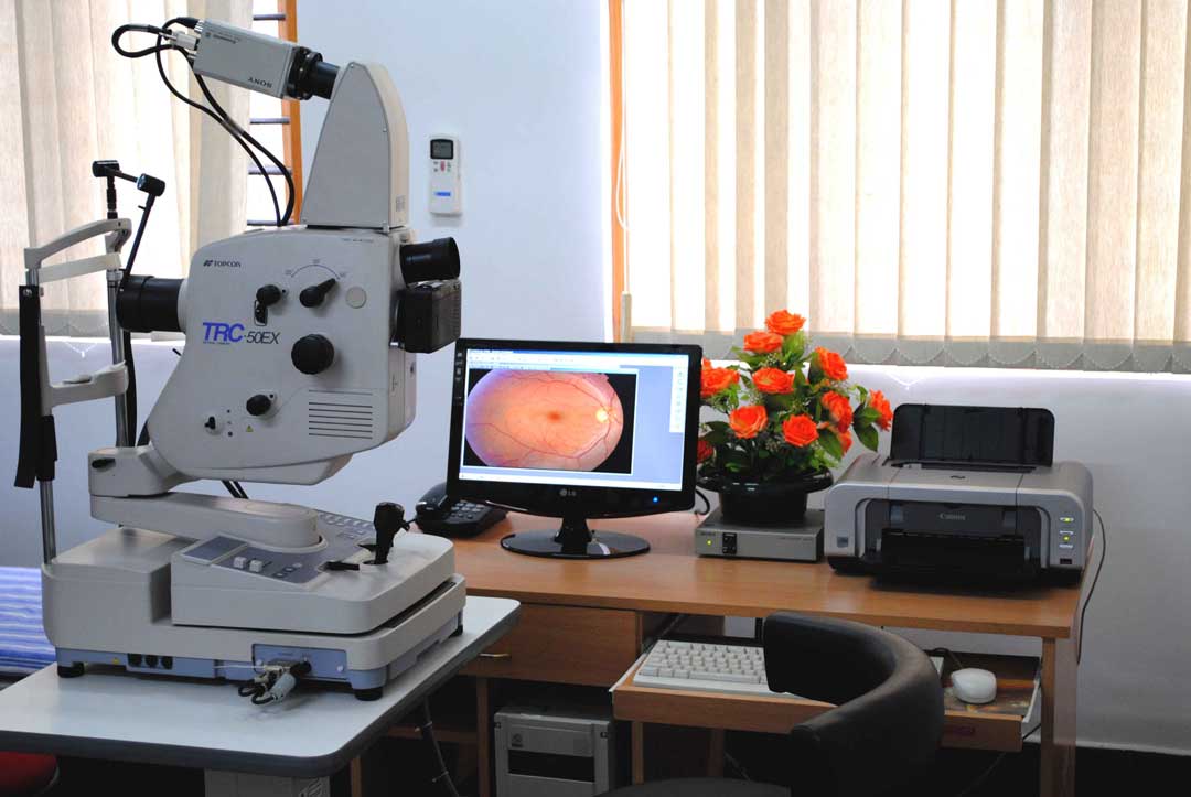
RETINAL IMAGING
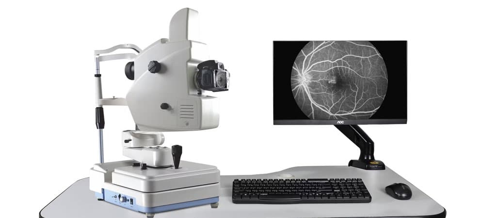
Fundus fluorescein angiography uses a yellow fluorescein dye to highlight retinal blood vessels, helping evaluate the back surface of the eye and diagnose conditions such as macular degeneration and diabetic retinopathy.
Fundus fluorescein angiography is a common procedure performed to evaluate the back surface of a patient's eye. A small amount of yellow fluorescein dye is injected into a vein in the patient's arm. The dye travels to the eye, where it highlights the blood vessels.
It helps diagnose eye disorders such as macular degeneration and diabetic retinopathy.
Macular Degeneration
Macular degeneration occurs in the macula, which is the part of the eye that allows you to focus on fine detail. Sometimes the disorder worsens so slowly that you may not notice any change at all. In some people, it causes vision to deteriorate rapidly, and blindness in both eyes may occur.
Because the disease destroys focused central vision, it can affect:
Diabetic Retinopathy
Diabetic retinopathy is caused by long-term diabetes and results in permanent damage to the blood vessels in the back of the eye, or the retina. The retina converts images and light that enter the eye into signals, which are then transmitted to the brain through the optic nerve.
There are two types of this disorder:
Fundus fluorescein angiography is used to determine if treatments for these eye disorders are working.
Normal Results
If the eyes are healthy, the blood vessels will have normal shape and size. There will be no blockages or leaks in the vessels.
Abnormal Results
Abnormal results may reveal a leak or blockage in the blood vessels. This may be due to: