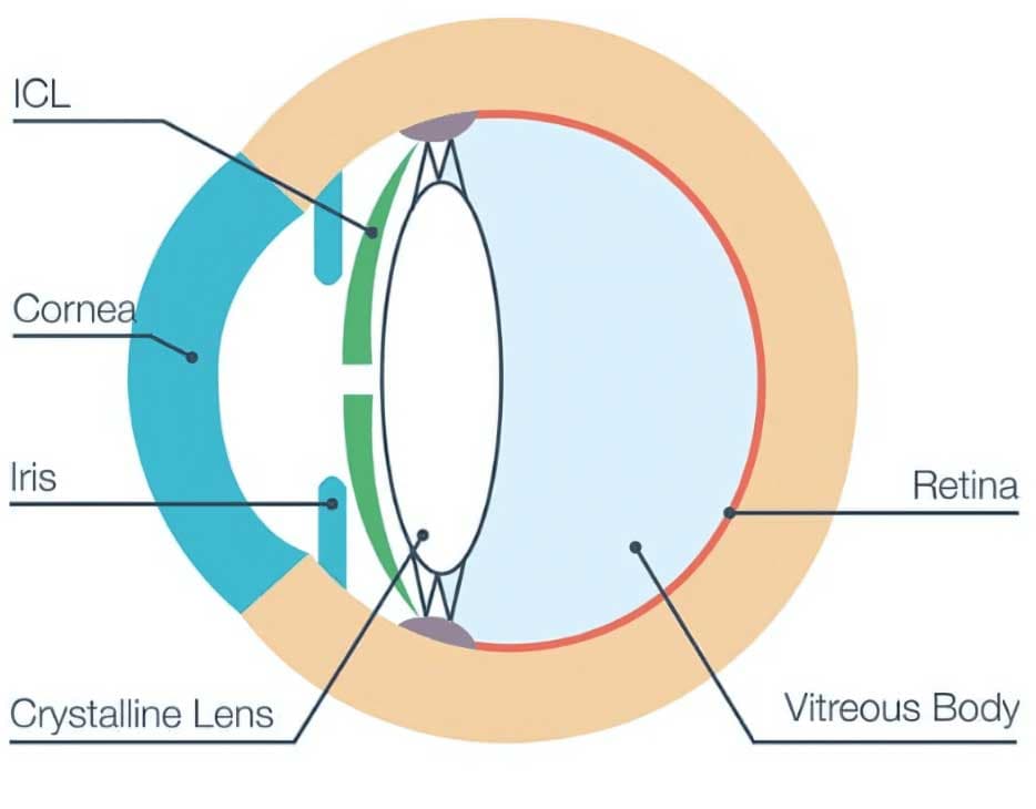
Precision-Led ICL Planning
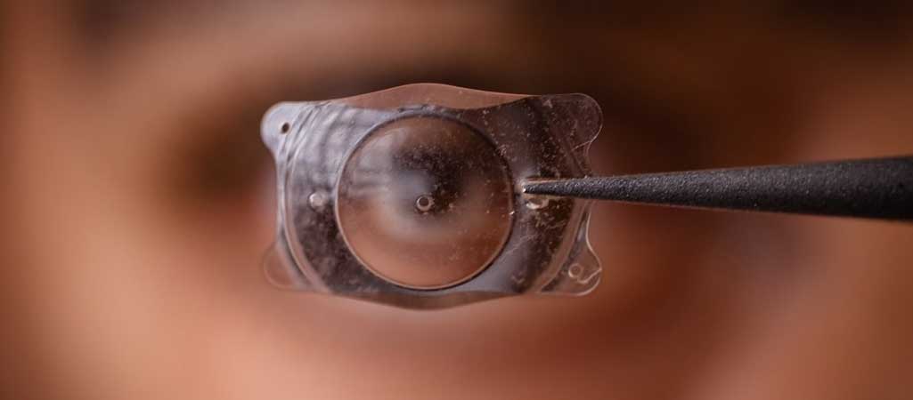
ICL surgery uses detailed eye measurements, a small incision, and careful lens placement to correct vision while preserving the cornea.
At Kenia Eye Hospital, ICL surgery is planned around precise diagnostics and individual eye anatomy. The aim is to correct the way light focuses on the retina without creating a corneal flap or reshaping the cornea.
Before surgery, measurements such as anterior chamber depth, white-to-white distance, sulcus sizing, and vault planning are reviewed. These help select a lens that fits the patient's eye anatomy and supports stable visual results.
Anesthetic eye drops are used to numb the eye for comfort. Because ICL does not require a corneal flap, the natural corneal structure and tear film are preserved.
A tiny corneal incision is made, and a foldable posterior chamber phakic intraocular lens is inserted. The lens is positioned between the iris and the natural lens, where it works to focus light clearly.
Once the ICL is placed, the surgeon checks optical alignment, lens position, and vault. The incision usually seals naturally without stitches.
Patients return home the same day after a short observation period. Follow-ups are used to monitor healing, intraocular pressure, lens position, endothelial health, and long-term outcomes.